Immunohistochemistry (IHC) is a technique that utilizes the principle of specific binding between antigens and antibodies in immunology. It identifies antigens (such as polypeptides and proteins) in tissue cells through chemical reactions that cause color-developing agents (fluorescein, enzymes, metal ions, isotopes) labeled on antibodies to develop color, enabling their localization, qualitative, and quantitative study. Due to its strong specificity, high sensitivity, and rapid and simple operation, immunofluorescence technology is widely applied in clinical pathological diagnosis and testing.
The IHC experimental process typically includes the following steps: tissue sample fixation, paraffin embedding, sectioning, dewaxing and hydration of sections, antigen retrieval, inactivation, blocking, primary antibody incubation, secondary antibody incubation, staining and color development, counterstaining, dehydration, coverslipping, and observation.
IHC staining Principle
Tissue Sample Acquisition and Fixation
Obtain the target tissue sample from animals or humans, and promptly (usually within 30 minutes after excision) place it into a fixative (commonly 10% neutral buffered formalin, methanol, ethanol, or their mixtures) with a volume 10-20 times that of the tissue to ensure the tissue is fully immersed in the fixative. The fixation time varies depending on tissue size and type, typically 18-24 hours.
Principles:
- Preserve Structure: Fixatives (such as formaldehyde) cross-link proteins to rapidly terminate intracellular enzyme activity, prevent tissue autolysis and decay, and maximize the preservation of tissue cell morphology and antigenicity.
- Stabilize Antigens: Fixation stabilizes antigen molecules in their original tissue positions, preventing diffusion or loss during subsequent processing.
- Harden Tissue: It hardens the tissue, facilitating subsequent sectioning operations.
Dehydration, Clearing, and Wax Infiltration
Dehydration: Pass the fixed tissue sample through gradient ethanol for thorough dehydration (70%, 80%, 95%, absolute ethanol 1, absolute ethanol 2, each soaking for 1 minute) → Clearing Agent: Use xylene to replace ethanol and make the tissue transparent → Wax Infiltration: Place the transparent tissue sample into melted paraffin. Sufficient time is required for each step to allow the reagent to fully penetrate the tissue.
Principles:
- Dehydration: Removes water from the tissue because water is incompatible with paraffin. Ethanol is a commonly used dehydrating agent.
- Clearing: Replaces ethanol with a clearing agent (such as xylene). The clearing agent can mix with ethanol and dissolve paraffin, serving as an intermediate medium.
- Wax Infiltration: Molten paraffin penetrates into the tissue to replace the clearing agent. After cooling, the paraffin solidifies, providing support for the tissue to facilitate cutting into thin sections.
Embedding and Slicing
The paraffin-impregnated tissue block is placed into an embedding mold, melted paraffin is added, and cooled and solidified into a wax block. The wax block was cut into 4-5 μm thick continuous slices using a slicer. Sections were floated in a warm water bath (40-45℃ ) to spread, then carefully fished onto slides (usually using anti-dehiscence slides such as poly-lysine or APES-treated slides) and baked in an oven at 60℃ for 2h.
Principles:
- Embedding: to form regular, firm wax blocks for fixation and sectioning.
- Sectioning: to obtain a layer of tissue thin enough to transmit light and be viewed under a microscope.
- Baking: slightly melts the paraffin wax in the section, better adheres to the slide and prevents the section from falling off during subsequent processing.
Dewaxing and Hydration
Place the glass slides sequentially into xylene I → xylene II (or substitutes) → absolute ethanol → absolute ethanol → 95% ethanol → 80% ethanol → 70% ethanol → deionized water. Soak each step for sufficient time (usually 5-10 minutes) to ensure complete removal of paraffin, as residual paraffin will affect subsequent antigen detection and staining. Avoid drying throughout the process, as drying can lead to non-specific antibody binding and high background staining.
Principles:
- Dewaxing: Dissolves and removes the paraffin embedding the tissue using xylene.
- Hydration: Gradually replaces xylene with decreasing concentrations of ethanol and finally replaces ethanol with water to restore the tissue to a hydrated state, facilitating the penetration and reaction of subsequent aqueous reagents (such as buffers and antibodies).
Antigen Retrieval
Common antigen retrieval methods include high-pressure heat retrieval, microwave heat retrieval, water bath heat retrieval, and trypsin retrieval. Typically, 1× sodium citrate (pH6.0) is used as the retrieval solution. For high-pressure retrieval, place the sections in a pressure cooker, add the antigen retrieval solution, heat until steaming, continue heating for 5 minutes, turn off the heat, and cool to room temperature to complete the retrieval (different antigens are suitable for different antigen retrieval solutions and conditions, and the most appropriate antigen retrieval method needs to be selected according to specific experimental circumstances).
Principles:
During formaldehyde or paraformaldehyde fixation, some antigens in the tissue undergo protein cross-linking and aldehyde group closure, losing their antigenicity. Antigen retrieval re-exposes intracellular antigenic determinants, improving antigen detection rates
Inactivation
Inactivate Peroxidase: For HRP enzyme-labeled systems, incubate the sections with 3% hydrogen peroxide aqueous solution at room temperature for 10-15 minutes (hydrogen peroxide is prepared fresh and stored at 4°C in the dark).
Principles:
Certain tissues (such as red blood cells, liver, kidney, and granulocytes) contain endogenous peroxidase/alkaline phosphatase or biotin, which can cause non-specific background staining. This step aims to inactivate or block these endogenous substances, reduce background signals, and improve the specificity and sensitivity of the experiment.
Blocking
Incubate the sections with a buffer (such as TBS or PBS) containing proteins (such as 5-10% normal serum - usually from the host animal of the secondary antibody, or 1-5% BSA) at room temperature for 10-30 minutes, but over-blocking should also be prevented. Blocking can also use calf serum, BSA, sheep serum, etc., but it must not be consistent with the source of the primary antibody.
Principles:
There are remaining sites on the tissue sections that can non-specifically bind to the primary antibody, causing false-positive results in subsequent steps. The proteins (serum or BSA) in the blocking solution will preemptively occupy these non-specific binding sites, preventing subsequent antibodies from non-specifically adsorbing here, thereby significantly reducing background staining.
Primary Antibody Incubation
Dilute the antibody with an antibody diluent and shake off the blocking solution on the slides. Primary antibody incubation is generally performed at 37°C for 1-2 hours or overnight at 4°C. Specific conditions still need to be optimized. After incubation, thoroughly wash the sections with a buffer (TBS or PBS, usually containing 0.05-0.1% Tween-20 and other mild detergents), typically 3 times for 5 minutes each time.
Principles:
Removes unbound primary antibodies or those non-specifically adsorbed on the tissue sections to reduce background staining. The detergent in the washing buffer helps remove loosely bound antibodies.
Secondary Antibody Incubation
Incubate the secondary antibody at room temperature for 30-60 minutes. After incubation, thoroughly wash with washing buffer 3 times for 5 minutes each time to remove unbound secondary antibodies.
DAB Color Development
Add freshly prepared DAB color development solution to the tissue. Closely monitor the color development reaction under a microscope (usually for seconds to tens of minutes at room temperature). When specific brown signals are clearly visible and the background is still low, immediately immerse the sections in deionized water to terminate the reaction.
Principles:
Enzymes (HRP or AP) catalyze specific substrate reactions to generate insoluble colored precipitates (color development method), which deposit at the antigen-antibody binding sites, thereby visualizing the location of the target antigen under an optical microscope. The color development time is critical; excessive time will lead to increased background and even non-specific staining.
Hematoxylin is a basic dye that mainly stains the chromatin (rich in DNA, negatively charged) of the cell nucleus blue. Counterstaining is used to form cell contours, helping to locate the position of the target protein (such as brown DAB-positive) within the cell and observe the tissue structure.
Counterstaining
Immerse the color-developed sections in hematoxylin stain for seconds to minutes (adjust according to the stain concentration and room temperature), differentiate (remove excess dye with acidic ethanol or water), and blue (rinse with tap water to make the cell nucleus blue).
Principles:
Hematoxylin is a basic dye that mainly stains the chromatin (rich in DNA, negatively charged) of the cell nucleus blue. Counterstaining is used to form cell contours, helping to locate the position of the target protein (such as brown DAB-positive) within the cell and observe the tissue structure.
Dehydration, Clearing, and Coverslipping
Dehydration: Place the slides sequentially into 70% ethanol → 80% ethanol → 95% ethanol → absolute ethanol → absolute ethanol → xylene → xylene (or substitutes). Soak each step briefly (usually 1-3 minutes). Remove the sections from xylene, add an appropriate amount of neutral gum (such as DPX) or aqueous mounting medium (such as those used for fluorescence or certain water-soluble color development products) to the tissue area, and carefully cover with a coverslip to avoid air bubbles.
Principles:
Dehydration: Removes water from the tissue because water is incompatible with paraffin. Ethanol is a commonly used dehydrating agent.
Clearing: Replaces ethanol with a clearing agent (such as xylene). The clearing agent can mix with ethanol and dissolve paraffin, serving as an intermediate medium.
Wax Infiltration: Molten paraffin penetrates into the tissue to replace the clearing agent. After cooling, the paraffin solidifies, providing support for the tissue to facilitate cutting into thin sections.
Microscopic Observation and Result Analysis
After the mounting medium dries and solidifies, observe the sections under an optical microscope (bright field for color development methods, fluorescence microscope for fluorescence methods). Interpret and analyze based on the distribution (cellular localization: nucleus/cytoplasm/membrane), intensity (weak/moderate/strong), scope, and pattern (diffuse/focal/punctate, etc.) of the positive signals. Pay attention to comparison with negative and positive controls.
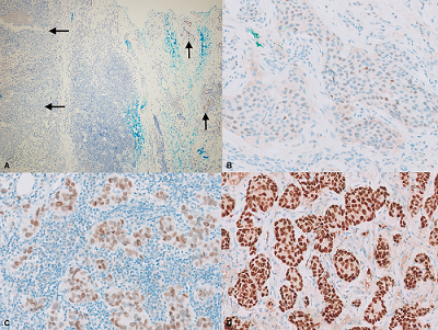
Immunohistochemistry for estrogen receptor in invasive breast carcinoma
(Arch Pathol Lab Med. 2022 Nov 1;146(11):1303-1307)
IHC FAQ
Non-Specific Staining?
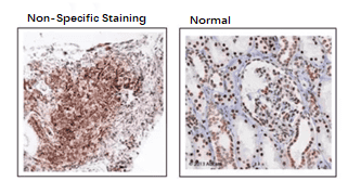
- Antibody Cross-Reactivity: Antibodies may bind to multiple antigens or non-specific structures in the sample, leading to non-specific staining. Selecting antibodies with high specificity, performing pre-absorption, or using competitive inhibition experiments can reduce this problem.
- Too High Antibody Concentration: Excessive antibody increases the chance of non-specific binding. Optimize the antibody concentration to the optimal range, usually determined through pre-experiments.
- Improper Sample Handling: Excessive fixation, dehydration, or solvents used during sample processing may expose additional binding sites, promoting non-specific binding. Optimize sample processing steps, use mild fixatives, and avoid excessive processing.
- Inadequate Washing: Inadequate washing steps fail to effectively remove unbound antibodies or markers, leading to non-specific staining. Increasing the number of washes or prolonging the washing time can improve this.
- Too High Sensitivity of the Detection System: An overly sensitive detection system may amplify non-specific signals. Adjust the sensitivity of the detection system or signal amplification steps to reduce non-specific staining.
- Endogenous Enzyme Activity: In enzyme-labeled antibody detection systems, the activity of endogenous enzymes (such as peroxidase) may cause non-specific staining. Using endogenous enzyme inhibitors or performing enzyme inactivation steps can reduce this effect.
- Complex Sample Background: Samples with high backgrounds may contain multiple potential binding sites, increasing the possibility of non-specific staining. Using blocking steps (such as serum or protein blocking solutions) can reduce background staining.
False Positives?
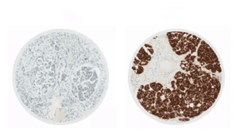
- Primary Antibody Concentration and Validity: Antibodies have an appropriate concentration range and must be used within the valid period. Expired antibodies may either fail to stain or cause background staining, forming false positives.
- Inadequate Reagent Coverage of Tissue: Reagents at the tissue edges are prone to drying, resulting in higher concentrations and deeper staining than in the middle of the tissue.
- Excessive Antibody Incubation Time: Due to temperature effects, incubation time should be slightly shorter in summer and slightly longer in winter.
- Tissue Drying During Operation: Tissue drying causes edge shrinkage or damage, forming artifacts and producing false positives.
- Too Long DAB Color Development Time: DAB should be prepared fresh, preferably within 30 minutes of preparation.
- Due to the high protein content in the cytoplasm, a lot of non-specific staining appears in the stroma and cytoplasm, such as the staining caused by endogenous enzymes hemoglobin, myoglobin, etc., which can be solved by serum blocking.
- Adipocytes and lymphocytes in breast cancer tissue can also produce non-specific staining.
- Non-specific antibody adsorption is common in necrotic tissue, causing high background staining in necrotic tissue. In immunohistochemistry, wax blocks with too much necrotic tissue should be avoided.
- Inadequate PBS rinsing, residual antibody results in enhanced staining.
Uneven Staining?
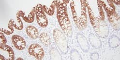
- Inconsistent Sample Preparation: Uneven section thickness or differences in sample fixation conditions lead to uneven distribution of the staining agent on the sample.
- Uneven Antibody Distribution: Uneven coverage of the antibody solution on the sample or insufficient penetration into all areas of the sample during incubation.
- Inadequate Antigen Retrieval: Inconsistent antigen retrieval conditions (such as temperature and time) lead to insufficient exposure of antigenic epitopes in some areas.
- Inadequate Washing Steps: Residual unbound antibodies or markers may accumulate in certain areas, causing excessively deep staining.
- Uneven Application of Color Developer: Uneven distribution of the color developer on the sample or inconsistent color development reaction time.
- Differences in Internal Sample Structure: Differences in internal sample structure (such as cell density and tissue type) may lead to different reactivity of the staining agent in different areas.
Weak Positivity?
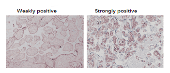
- Improper Fixation or Too High Fixation Temperature: Affects the quantity and quality of antigens.
- Inappropriate Antigen Retrieval Method: Insufficient exposure of antigenic determinants.
- Too High Antibody Dilution or Inadequate Incubation Temperature/Time:
- Excessive Rinse Solution Remaining on the Slides Before Antibody Incubation:
Slides Not Placed Horizontally During Incubation: Causing uneven antibody incubation
Partial List of Products Applicable to IHC
|
Catalog No. |
Product Name |
Applications |
|
Anti-MMP9 Polyclonal Antibody |
WB, IHC, IF |
|
|
Anti-BDNF Polyclonal Antibody |
WB, IHC, IF |
|
|
Anti-Human CD3E Polyclonal Antibody |
WB, IHC, IF |
|
|
Anti-VASN Polyclonal Antibody |
WB, IHC, IF |
|
|
Anti-Human Fibrin Antibody (59D8) |
WB, IHC, IF |
|
|
Anti-Human GZMB Antibody (SAA0516) |
WB, IHC, IF |
|
|
Anti-Human EDB/EDB-FN Antibody (SAA1375) |
WB, IHC, IF |
|
|
Anti-H3K4me3 Antibody (304M3-B) |
WB, IHC, IF |
|
|
Anti-Human VEGFA Antibody (SAA0531) |
WB, IHC, IF |
|
|
Anti-Human SULT1E1 Antibody (SAA0758) |
WB, IHC, IF |
|
|
Anti-Mouse CD68/Macrosialin Antibody (SAA2218) |
WB, IHC, IF |
|
|
Anti-Mouse CD229/LY9 Antibody (D08) |
WB, IHC, IF |
