
Japan Reports Over 11,600 HFMD Cases: What Are EV-71, CV-A16, and CV-A6?
Recent reports of Hand, Foot, and Mouth Disease (HFMD) in Japan have drawn increasing public health attention. According to the Japan Institute for Health Security (JIHS), the cumulative number of HFMD cases nationwide has exceeded 11,600 in 2026. During the week of May 25--31 alone, six prefectures, including Fukuoka, Miyazaki, and Kagoshima, reported case numbers above the official alert threshold, prompting local health authorities to issue outbreak warnings earlier than in previous years.
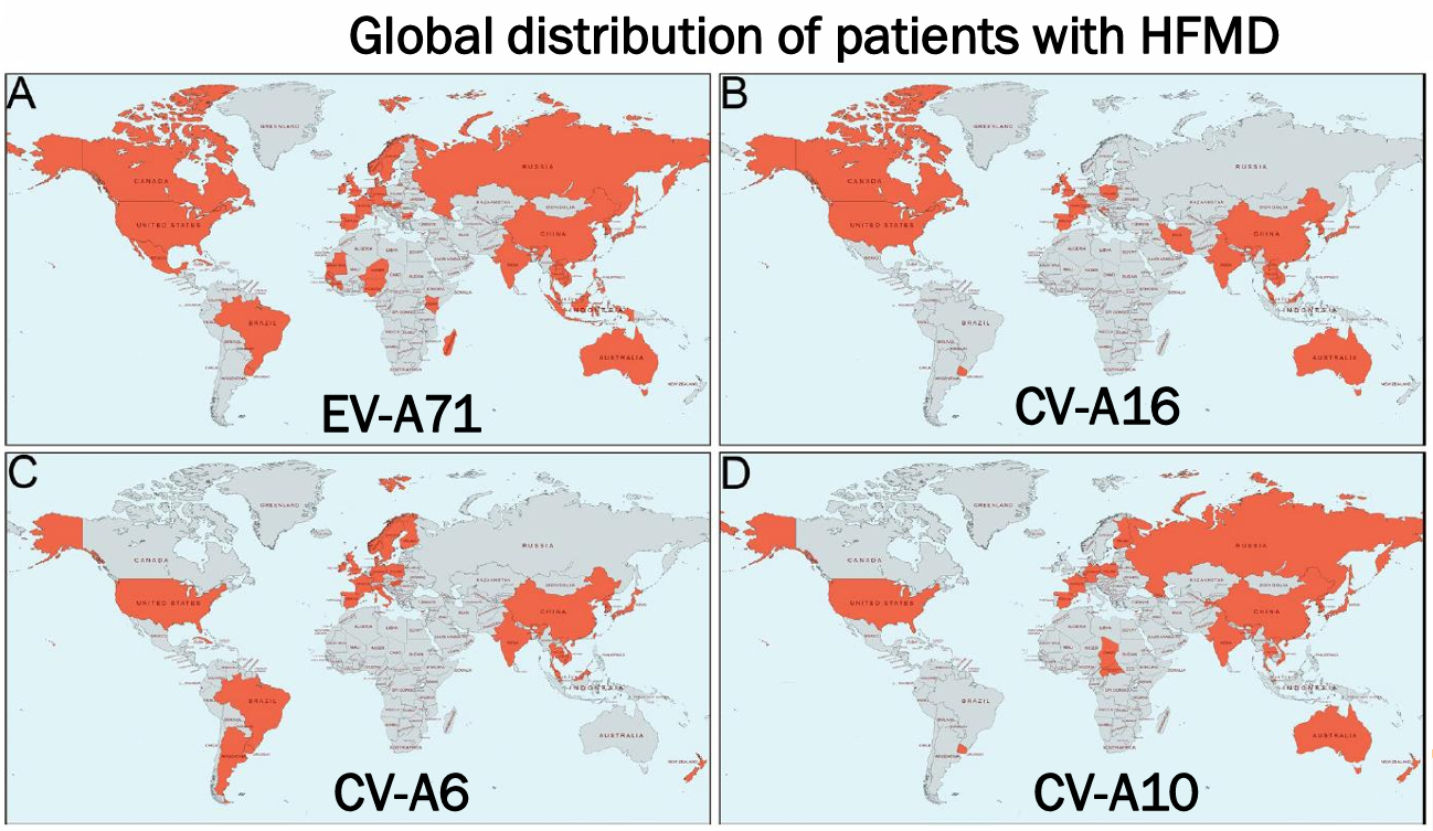
Global distribution of patients with HFMD
HFMD is one of the most common acute viral infections in young children. Driven by the continual emergence and replacement of circulating enterovirus serotypes, outbreaks occur regularly across the globe. The disease remains endemic in many regions and has caused recurrent outbreaks throughout North America, Europe, and the Asia-Pacific. Although most infections are self-limiting, the high transmissibility of HFMD, the diversity of causative pathogens, and the potential for severe neurological complications in a subset of patients continue to make it a significant public health concern.
In this article, we take a closer look at the major viral pathogens responsible for HFMD, covering their classification, genomic organization, pathogenic mechanisms, and current prevention strategies.
Major HFMD Pathogens
HFMD is caused by a diverse group of enteroviruses, with more than 20 serotypes reported to be associated with disease. Among them, Enterovirus A71 (EV-A71), Coxsackievirus A16 (CV-A16), Coxsackievirus A6 (CV-A6), and Coxsackievirus A10 (CV-A10) are the most clinically relevant.
Historically, EV-A71 and CV-A16 have been the predominant causative agents of HFMD. However, the epidemiological landscape has evolved considerably over the past decade. In many countries and regions, CV-A6 has emerged as a dominant circulating strain and, in some outbreaks, has surpassed both EV-A71 and CV-A16 in prevalence.
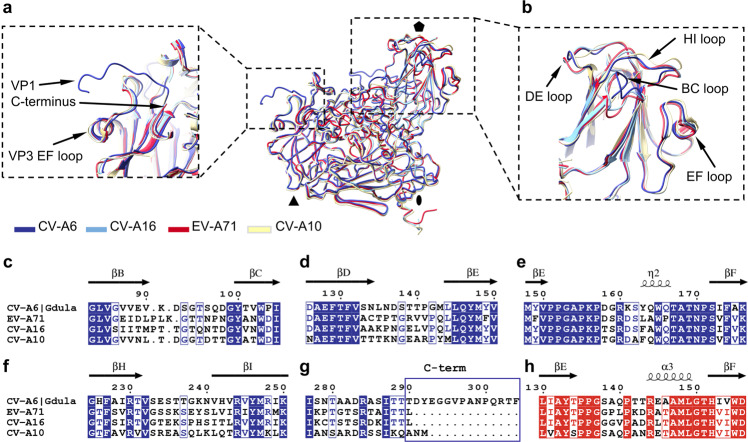
Comparison of structures of capsid protomers from virions of CV-A6, CV-A16, EV-71, and CV-A10 (PMCID: PMC9438360)
Clinical manifestations vary substantially among enterovirus serotypes. EV-A71 is the pathogen most strongly associated with severe HFMD, neurological complications, and fatal outcomes. Infections often present with persistent fever and can progress to meningitis, encephalitis, or brainstem encephalitis. In contrast, CV-A16 infections are generally milder and rarely involve the central nervous system.
Although CV-A6 exhibits lower neurovirulence than EV-A71, it is frequently associated with more extensive and atypical skin lesions. Rashes may extend beyond the hands, feet, and oral cavity, and patients commonly experience skin peeling and nail shedding during recovery, creating additional challenges for clinical diagnosis.
Genome Organization and Virion Structure
EV-A71 and CV-A16 belong to the genus Enterovirus within the family Picornaviridae. Both are non-enveloped viruses with an icosahedral capsid approximately 20--30 nm in diameter.
Their genomes consist of a positive-sense single-stranded RNA molecule of approximately 7.4--7.5 kb. The genome contains a single open reading frame (ORF) that is translated into a polyprotein precursor. Viral proteases subsequently process the polyprotein into three precursor regions: P1, P2, and P3.
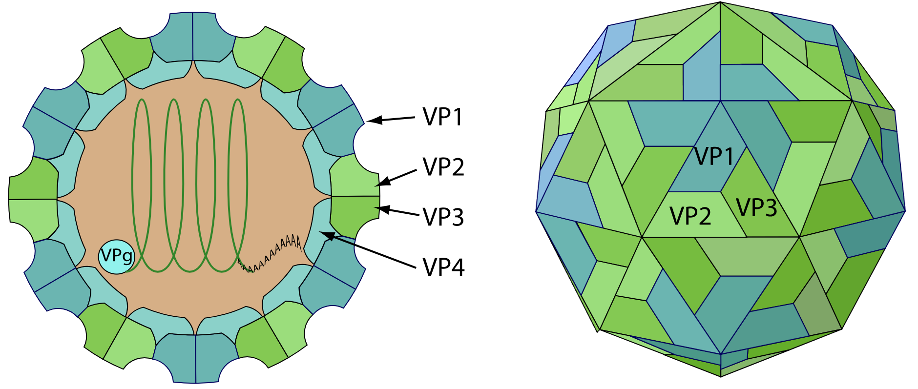
Enterovirus virion structure
The P1 region gives rise to four structural proteins---VP1, VP2, VP3, and VP4---which collectively form the viral capsid. The P2 and P3 regions generate seven non-structural proteins involved in viral replication, polyprotein processing, and host-cell manipulation.
Structural Proteins
The enterovirus capsid is composed of 60 copies each of VP1, VP2, VP3, and VP4.
Among these proteins, VP1 is exposed on the virion surface and plays a critical role in receptor recognition, including binding to SCARB2, one of the major receptors for EV-A71 infection. VP1 also contains the principal neutralizing epitopes and is therefore a primary target for vaccine development, serological assays, and therapeutic antibody discovery.
VP2 and VP3 contribute to capsid integrity and antigenic structure, while VP4 is located internally and participates in viral uncoating and genome release during infection.
| Protein | Primary Function |
|---|---|
| VP1 | Receptor binding and major neutralizing antibody target |
| VP2 | Capsid stability and antigenic epitope formation |
| VP3 | Structural integrity and capsid assembly |
| VP4 | Viral uncoating and genome release |
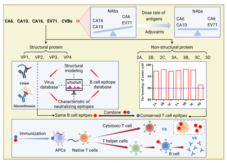
A series of potential methods to get more balanced immune responses and a broader spectrum of protective immune responses in development of multivalent HFMD vaccines. (PMCID: PMC9820767)
Non-Structural Proteins and Viral Pathogenesis
Non-structural proteins orchestrate virtually every step of the viral replication cycle. In addition to processing viral polyproteins and replicating viral RNA, they help the virus evade host defenses by suppressing cellular protein synthesis and interfering with innate immune responses.
The 2A and 3C proteases cleave viral polyproteins while simultaneously disrupting host cellular functions. The 2B protein alters membrane permeability, facilitating virus release, whereas 2C participates in the formation of replication organelles. VPg (3B) serves as a primer for RNA synthesis, and the RNA-dependent RNA polymerase 3D is responsible for genome replication and progeny virus production.
| Protein | Primary Function |
|---|---|
| 2A Protease | Polyprotein processing and host translation shutoff |
| 2B | Membrane permeabilization and virus release |
| 2C | ATPase/NTPase involved in replication organelle formation |
| 3A | Inhibition of intracellular protein trafficking |
| VPg (3B) | Primer for viral RNA synthesis |
| 3C Protease | Polyprotein processing and suppression of host responses |
| 3D Polymerase | RNA-dependent RNA polymerase responsible for genome replication |
Through the coordinated activities of structural and non-structural proteins, enteroviruses complete a complex life cycle encompassing attachment, entry, uncoating, translation, genome replication, capsid assembly, and viral release.
Diagnosis and Prevention
Laboratory diagnosis of HFMD relies on both molecular and serological approaches.
Reverse transcription PCR (RT-PCR) remains the gold standard for pathogen detection and genotyping, particularly through amplification and sequencing of the VP1 region. Serological methods such as ELISA are widely used to detect virus-specific IgM and IgG antibodies for infection confirmation and epidemiological surveillance. In recent years, antigen detection assays targeting multiple enterovirus serotypes have also been developed.
Currently, no specific antiviral therapy is available for HFMD. Inactivated EV-A71 vaccines have been licensed in China and have demonstrated strong protection against EV-A71-associated disease. However, due to the extensive serotype diversity of enteroviruses and the limited cross-protection among them, monovalent EV-A71 vaccines do not provide effective protection against CV-A6, CV-A16, or other circulating serotypes.
As CV-A6 and CV-A10 continue to rise in prevalence, the development of multivalent vaccines capable of providing broad protection against multiple HFMD-associated enteroviruses has become an important research priority.
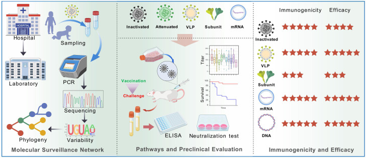
HFMD vaccine development strategies (PMCID: PMC12179169)
AntibodySystem Supports HFMD Research
Reliable detection tools and high-quality research reagents are essential for advancing HFMD research and diagnostic development.
AntibodySystem offers a comprehensive portfolio of recombinant proteins, research-grade antibodies, and ELISA kits covering major HFMD-associated enteroviruses, including EV-A71, CV-A16, CV-A6, and CV-A10.
These products support a broad range of applications, including viral pathogenesis studies, host immune response characterization, antibody discovery, vaccine research, and diagnostic assay development.
Human enterovirus 71, EV71
| Catalog | Product Name |
|---|---|
| YVV20602 | Recombinant EV71 P3C/Protease 3C Protein, N-His |
| YVV20601 | Recombinant EV71 Protease 3C Protein, C-His |
| YVV20501 | Recombinant EV71 VP3/Capsid protein VP3 Protein, N-GST |
| YVV20401 | Recombinant EV71 VP0/Capsid protein VP0 Protein, N-GST |
| YVV20303 | Recombinant EV71 VP4/P1A Protein, N-GST & C-His |
| YVV20302 | Recombinant EV71 P2A/Protease 2A Protein, N-His |
| YVV20301 | Recombinant EV71 VP1/Capsid protein VP1 Protein, N-His |
| VVV20304 | InVivoMAb Anti-EV71 Mature virion in Complex Antibody (D6) |
| VVV20303 | InVivoMAb Anti-EV71 Mature virion in Complex Antibody (A9) |
| RVV20303 | Anti-EV71 Capsid protein VP1/2/3 Antibody (SAA2027) |
| PVV20401 | Anti-EV71 VP0/Capsid protein VP0 Polyclonal Antibody |
| PVV20301 | Anti-EV71 VP1/Capsid protein VP1 Polyclonal Antibody |
| KAV20301 | Anti-EV71 Capsid protein VP1 hIgG ELISA Kit |
Coxsackievirus, CVA
| Catalog | Product Name |
|---|---|
| YVV20702 | Recombinant CV-A10 VP1/P1D Protein, N-GST/C-His |
| YVV19102 | Recombinant CVA6 VP2/Capsid protein VP2 Protein, N-His |
| YVV19101 | Recombinant CVA6 VP1/Capsid protein VP1 Protein, N-His |
| VVV21702 | InVivoMAb Anti-CVA16 mature virion in complex (Iv0109) |
| VVV21701 | InVivoMAb Anti-CVA16 mature virion in complex (Iv0108) |
| RVV19201 | Anti-Coxsackievirus A16/CVA16 Capsid protein/Genome polyprotein Antibody (SAA0355) |
| RVV19102 | Mouse Anti-Coxsackievirus A6/CVA6 VP1/Capsid protein VP1 Antibody (SAA0354) |
| RVV19101 | Anti-Coxsackievirus A6/CVA6 VP1/Capsid protein VP1 Antibody (SAA0354) |


