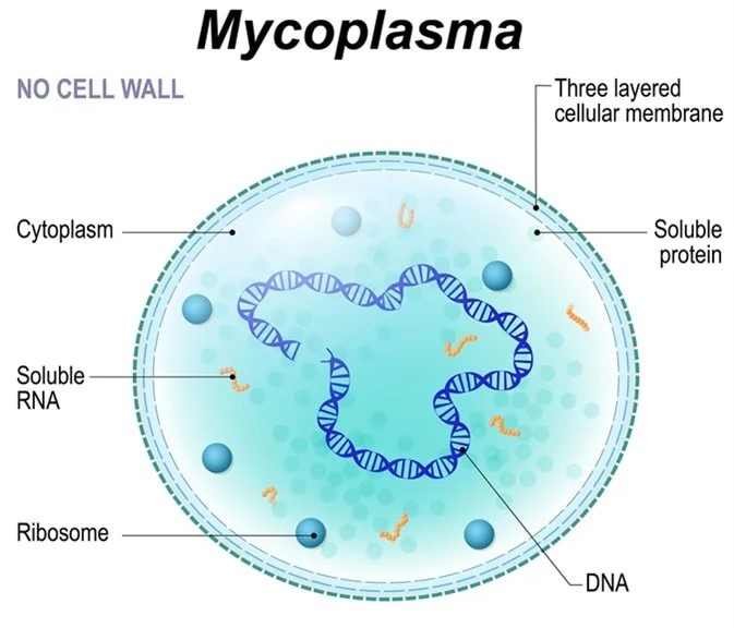
Mycoplasma pneumoniae (MP), one of the smallest free-living microorganisms known, occupies an evolutionary niche between bacteria and viruses. This single-celled organism measures 0.2–0.3 μm in diameter and 0.5–1.0 μm in length. Lacking a cell wall, it exhibits intrinsic resistance to penicillin and cephalosporin antibiotics. Its pleomorphic morphology varies with environmental conditions and growth stages. The MP cell comprises a plasma membrane, ribosomes, and double-stranded DNA, but lacks a nucleus, cell wall, flagella, or spores.Mycoplasma pneumoniae (MP), one of the smallest free-living microorganisms known, occupies an evolutionary niche between bacteria and viruses. This single-celled organism measures 0.2–0.3 μm in diameter and 0.5–1.0 μm in length. Lacking a cell wall, it exhibits intrinsic resistance to penicillin and cephalosporin antibiotics. Its pleomorphic morphology varies with environmental conditions and growth stages. The MP cell comprises a plasma membrane, ribosomes, and double-stranded DNA, but lacks a nucleus, cell wall, flagella, or spores.

Figure1. Schematic structure of Mycoplasma pneumoniae
Transmission Dynamics
MP demonstrates global distribution, causing pandemics every 2–6 years. While all populations are susceptible, MP pneumonia predominantly affects children and young adults. Outbreaks frequently occur in densely populated settings such as schools and hospitals.
As the leading cause of community-acquired pneumonia in China, MP invades respiratory epithelial cells through specialized adhesion mechanisms. Clinical manifestations range from upper respiratory infections to severe atypical pneumonia and diverse extrapulmonary complications. As an obligate human pathogen, MP spreads via respiratory droplets/aerosols from infected individuals or carriers. Transmission occurs during both incubation and treatment phases, with peak infectiousness within the first 4–6 days of illness, explaining high intrahousehold transmission rates.
Pathogenesis
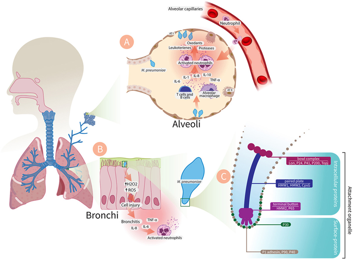
The entry of Mycoplasma pneumoniae into the alveolar space as a pathogen causes the activation of inflammatory reactions by immune cells, at the head of which are the alveolar macrophages. Macrophages cause the call of neutrophils and their migration from the alveolar capillaries by producing and releasing cytokines. Activated neutrophils in the alveolar space release leukotrienes, proteases and oxidants which increase tissue damage and alveolar edema. The adhesion of Mycoplasma pneumoniae to bronchial cells can cause oxidative stress and increase cell damage and cell death through the production of oxidants like H2O2, which can lead to bronchitis

Figure 2. Mycoplasma pneumonia Pathogenesis
(Microb Pathog. 2024 Nov:196:106944.)
MP pathogenicity hinges critically on its adhesion capacity, mediated by key virulence factors:
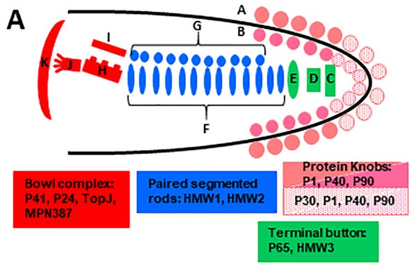
P1 protein: Primary 170-kDa adhesin (1,627 aa) localizing to the attachment organelle tip. Essential for host receptor binding, adhesion, gliding motility, and release of CARDS toxin, H₂O₂, and superoxide radicals.
P30 protein: 30-kDa membrane protein stabilizing the organelle tip, facilitating adhesion and gliding.
HMW proteins (HMW1-3): High-molecular-weight complex regulating P1 localization and organelle structure.
P65 protein: Core structural component enabling adhesion and motility.
CARDS toxin: Community-Acquired Respiratory Distress Syndrome toxin released via P1, inducing host cell damage.
P90/P40 proteins: Anchor P1 to the organelle cytoskeleton for functional stability.
These proteins collectively enable respiratory epithelial colonization, immune evasion, and infection establishment.

Figure 3. Schematic of M. pneumoniae terminal organelle architecture
(Mol Microbiol. 2018 May;108(3):306-318.)
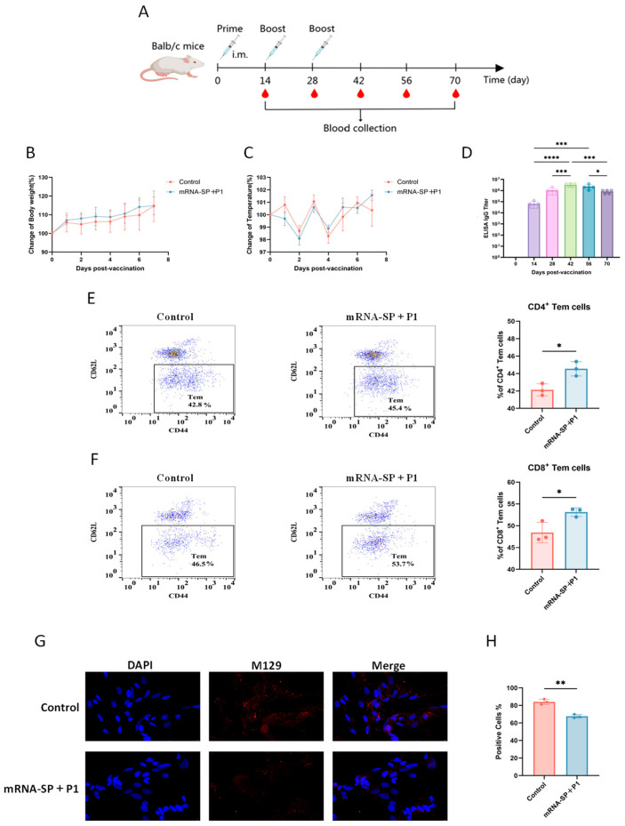
In July, a study published in the International Journal of Molecular Sciences under the title "An mRNA Vaccine Targeting the C-Terminal Region of P1 Protein Induces an Immune Response and Protects Against Mycoplasma pneumoniae" utilized AntibodySystem’s Recombinant Mycoplasma pneumoniae mgpA/Adhesin P1 Protein, N-His (catalog YXX09301) to evaluate the humoral immune response elicited by the vaccine. This recombinant P1 protein served as the coating antigen in ELISA to quantify P1-specific IgG antibody levels in sera from mice immunized with the mRNA-SP+P1 vaccine. Results demonstrated that the vaccine effectively induced high-titer antibodies (post-third-dose titers reaching 10⁶–10⁷), with confirmed functional activity: immune sera significantly inhibited M. pneumoniae adhesion to host cells.

References
1. Georgakopoulou V, Ioannis Lempesis, Pagona Sklapani, Trakas N, Demetrios Spandidos. Exploring the pathogenetic mechanisms of Mycoplasma pneumoniae (Review). Experimental and Therapeutic Medicine. 2024;28(1).
2. Hu J, Ye Y, Chen X, Xiong L, Xie W, Liu P. Insight into the Pathogenic Mechanism of Mycoplasma pneumoniae. Current Microbiology. 2022;80(1).
3. Hamed Tahmasebi, Babaeizad A, Mohammadlou M, Farnaz Alibabaei, Seyedeh Zahra Banihashemian, Eslami M. Microbial Pathogenesis. 2024;196:106944-106944.
4. Jiang Z, Li S, Zhu C, Zhou R, Leung PHM. Mycoplasma pneumoniae Infections: Pathogenesis and Vaccine Development. Pathogens. 2021;10(2):119.
5. Krause DC, Chen S, Shi J, Jensen AJ, Sheppard ES, Jensen GJ. . Molecular microbiology. 2018;108(3):306-318.
6. Zhang F, Li C, Wu Y, et al. An mRNA Vaccine Targeting the C-Terminal Region of P1 Protein Induces an Immune Response and Protects Against Mycoplasma pneumoniae. International Journal of Molecular Sciences. 2025;26(13):6536-6536.
MP-Related Products
Recombinant Protein
|
Catalog |
Product Name |
|
YXX09301 |
Recombinant Mycoplasma pneumoniae mgpA/Adhesin P1 Protein, N-His |
|
YXX16801 |
Recombinant Mycoplasma pneumoniae CARDS Protein, N-His |
|
YXX17401 |
Recombinant Mycoplasma pneumoniae P30 adhesin Protein, N-His |
|
YXX16601 |
Recombinant Mycoplasma pneumoniae 116 kDa surface antigen Protein, N-His |
|
YXX16701 |
Recombinant Mycoplasma pneumoniae HMW3 Protein, N-His-SUMO |
|
YXX16901 |
Recombinant Mycoplasma pneumoniae p65 Protein, N-His |
|
YXX09302 |
Recombinant Mycoplasma pneumoniae mgpA/Adhesin P1 Protein, N-His |
|
YXX17301 |
Recombinant Mycoplasma pneumoniae MP438/MPN400 Protein, N-His |
|
YXX09303 |
Recombinant Mycoplasma pneumoniae mgpA/Adhesin P1 Protein, N-His |
|
YXX17501 |
Recombinant Mycoplasma pneumoniae Mgp3/P90 Protein, N-His |
Antibody
|
Catalog |
Product Name |
|
PXX09301 |
Anti-Mycoplasma pneumoniae mgpA/Adhesin P1 Polyclonal Antibody |
|
PXX16801 |
Anti-Mycoplasma pneumoniae CARDS Polyclonal Antibody |
|
PXX17401 |
Anti-Mycoplasma pneumoniae P30 adhesin Polyclonal Antibody |
