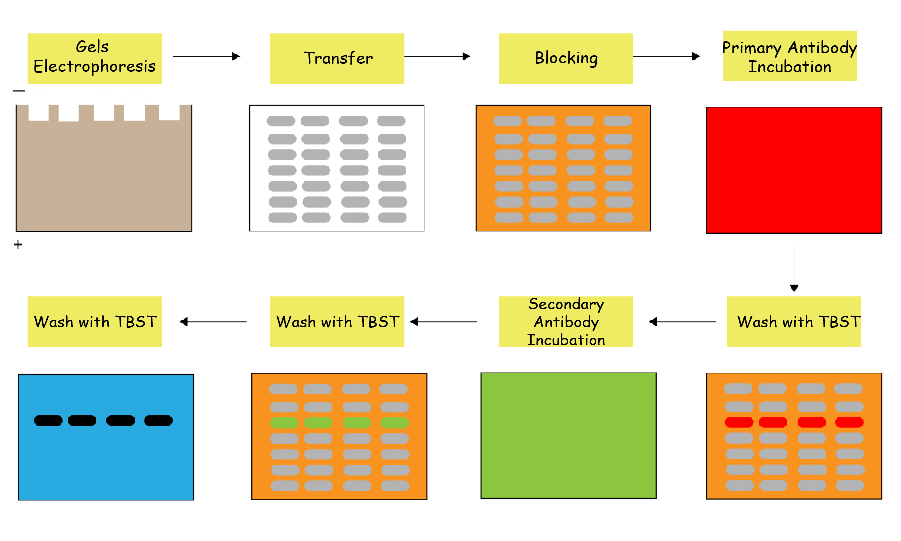
Weak bands, high background, inconsistent results... Who understands the pain of Phosphorylated Proteins Western Blot (WB)? The "nightmare" status of phosphorylated proteins WB stems from its inherent low abundance, high dynamism, and susceptibility to degradation. Minor operational deviations can cause signal loss or nonspecific amplification. Today, Dr. Connie analyzes these difficulties and shares actionable solutions.

Phosphorylated Proteins WB Difficulties & Solutions
Sample Preparation
·Low abundance: Phosphorylated proteinss typically constitute only 1%-10% of total cellular protein, pushing detection signals close to WB’s sensitivity limit.
·Narrow dynamic range: Kinase/phosphatase activity causes rapid phosphorylation fluctuations (e.g., EGFR phosphorylation peaks at 5 min post-EGF stimulation and returns to baseline by 30 min), making true signal capture timing-critical.
·Persistent phosphatase activity: Cell lysis releases endogenous phosphatases that catalyze rapid dephosphorylation, leading to signal decay.
·Temperature sensitivity: Phosphorylated proteins half-lives can be minutes at room temperature.
Thus, sample quality dictates success. Failures often originate here!
Critical steps
·Maintain ice-cold conditions throughout protein extraction.
·Supplement lysis buffers with phosphatase and protease inhibitors to prevent degradation/dephosphorylation.
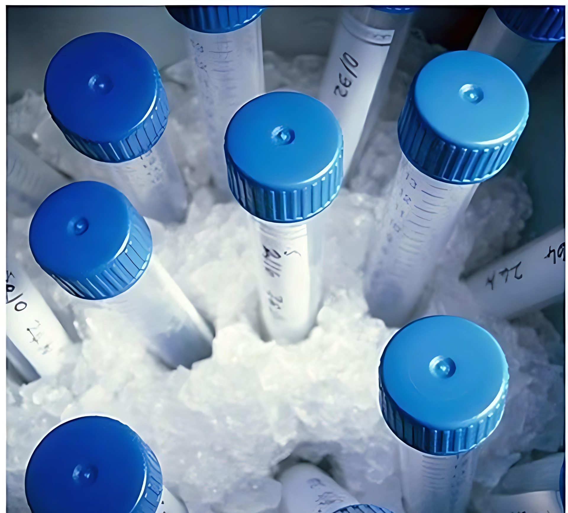
·Prioritize fresh samples flash-frozen in liquid nitrogen. Store at -80°C in aliquots; avoid freeze-thaw cycles.
·Load sufficient protein during electrophoresis.

Antibody Selection
·Consult high-impact literature to confirm phosphorylation sites. Use validated phospho-specific antibodies for enhanced sensitivity (batch variability exists; use consistent batches for comparable experiments).
·Total target protein serves as an internal control, distinguishing whether phosphorylation changes occur independently of total protein expression. Always pair phospho-target detection with total target protein detection (e.g., detect both p-Akt and Akt). Only phospho/total protein ratio changes confirm phosphorylation-specific regulation.
Electrophoresis & Transfer
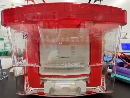
·Choose appropriate gel concentrations based on target molecular weight. Perform electrophoresis on ice to prevent thermal degradation.
·Phospho-WB frequently shows nonspecific bands. Use high-precision protein marker to confirm target size and avoid false positives.
·Prepare fresh transfer buffer to maximize efficiency.
·PVDF membranes outperform NC membranes in capturing phosphorylated proteinss and are recommended for phospho-protein transfer.
·Small phosphorylated proteinss may over-transfer. Empirically optimize transfer time/voltage (reduce time or voltage if needed).

Blocking & Antibody Incubation
·Blocking: Use 5% BSA (superior to 5% skim milk for many users). Some report high background and smearing with skim milk, potentially due to phosphorylated proteins casein in milk causing degradation. While some labs successfully use skim milk, optimize based on empirical results. Block at RT for 1h; avoid over-blocking.
·Antibody incubation: Dilute antibodies per manufacturer’s protocol or established literature. Incubate primary antibodies at 4°C overnight for optimal binding. Titrate antibody concentration and incubation time experimentally.
Every step in phospho-WB impacts outcomes. Prioritize preserving phosphorylation states and continuously optimize protocols for accuracy and reproducibility!
|
Catalog. |
Product Name |
|
RGK26901 |
Anti-Pan-pS/pSer/Phosphoserine Antibody (SAA0307) |
|
RGK60101 |
Anti-Pan-pY/pTyr/Phosphotyrosine Antibody (4G10) |
|
RHJ88704 |
Anti-Phospho-RIPK3 (pS199) Antibody (SAA0298) |
|
RMD96501 |
Anti-Phospho-AKT1 (pS473) Antibody (P8.H9) |
|
RHF62702 |
Anti-Phospho-Histone H3 (pT11) Antibody (N123/19) |
|
RHJ88701 |
Anti-Phospho-RIP3 (pS227) Antibody (P2.A11) |
|
RHC82407 |
Anti-Phospho-Tau (pS396/pS404) Antibody (SAA0294) |
|
RHH20001 |
Anti-Phospho-Smad2 (pT8) Antibody (P4.B9) |
