Fructose is ubiquitous in modern diets—from fruits and sweetened beverages to candies and baked goods. However, emerging research reveals a critical concern: excessive fructose consumption during early life may severely disrupt brain development. On June 11, 2025, a team led by Justin S.A. Perry at Memorial Sloan Kettering Cancer Center (MSKCC) published a landmark study in Nature titled "Early life high fructose exposure disrupts microglia function and impedes neurodevelopment". Their findings sound a critical alarm: Exposure to high-fructose environments during pivotal developmental windows (pregnancy and the neonatal period) directly impairs microglia—the brain’s essential "custodian" cells—through a GLUT5-dependent mechanism. This disrupts synaptic pruning and neuronal clearance, compromises neurodevelopment, and significantly elevates lifelong risks of cognitive deficits and anxiety-like behaviors.
The Epidemiologic Link
Prior studies correlate maternal high-fructose diets (e.g., sugary drinks, juices) or adolescent overconsumption with disrupted neurodevelopment and mood disorders (e.g., anxiety) in offspring. The new study identifies how this occurs.
Core Discovery: Fructose Paralyzes the Brain’s "Gardeners"
During critical phases of brain development, the nervous system undergoes a refined "pruning" process: eliminating superfluous neurons and synaptic connections to optimize neural circuitry—much like a gardener selectively trims branches to foster healthy growth. This essential function is executed by microglia, the brain’s resident immune cells and professional phagocytes of the central nervous system (CNS). In both mammals and humans, microglia mediate synaptic pruning and clear apoptotic neurons through efferocytosis, thereby removing redundant cellular elements and maladaptive wiring. Compromised microglial phagocytosis disrupts this clearance mechanism, leading to accumulation of uncleared apoptotic neurons—a pathological state with detrimental consequences for neurodevelopment.
Main Research Contents
1.Fructose Reduces Microglia Numbers & Induces a Dormant State:
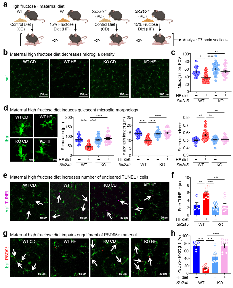
■Offspring from dams fed a clinically relevant high-fructose diet (15% kcal fructose, HF) showed significantly fewer microglia in the prefrontal cortex (PFC) vs. controls (CD) at postnatal day 7 (P7) (Fig. 1b, c).
■Remaining microglia exhibited morphological changes indicating dysfunction: reduced soma size, shorter major axis length, and increased roundness—phenotypes associated with a quiescent, hypo-phagocytic state (Fig. 1d).
■Consequences: Increased uncleared apoptotic cells (TUNEL+ nuclei, Fig. 1e, f) and impaired synaptic pruning (reduced microglial engulfment of PSD95+ material, Fig. 1g, h).

Fig. 1 Early life high fructose exposure suppresses microglial phagocytosis in vivo
2. The Culprit: GLUT5 (SLC2A5)-Dependent Fructose Transport:
■Microglia are the only CNS/immune cells expressing GLUT5, the high-affinity fructose transporter (Extended Data Fig. 3e,f).
■Strikingly, genetic deletion of Slc2a5 (GLUT5 KO) in neonates completely reversed HF-induced microglial loss, morphological alterations, and phagocytic defects (Fig. 1b-h).
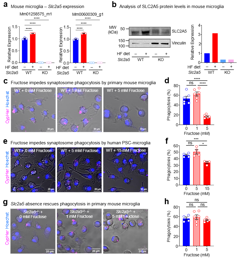
■In vitro, GLUT5-deficient primary microglia exposed to high fructose (5mM) showed normal phagocytosis of synaptosomes and apoptotic neurons, unlike impaired wild-type (WT) cells (Fig. 2g, h; Extended Data Fig. 5f).

Fig.2 High fructose exposure directly suppresses microglia phagocytosis
3.Metabolic Reprogramming Underlies Functional Impairment:
■High fructose directly suppressed phagocytosis in primary mouse microglia (Fig. 2c, d) and human iPSC-derived microglia (Fig. 2e, f).
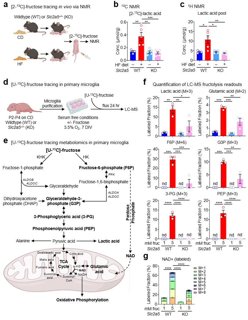
■Metabolic tracing ([U-¹³C]-fructose LC-MS / [2-¹³C]-fructose NMR) revealed GLUT5-dependent fructose uptake and catabolism (fructolysis) in neonatal brains (Fig. 3b, c) and microglia.
■HF-exposed microglia exhibited increased flux into fructose-6-phosphate (F6P) via hexokinase (HK) and a shift toward an oxidized metabolic state (↓ reduced glutathione, ↑ NAD⁺/NADH ratio) (Fig. 3f, g; Extended Data Fig. 6c)—phenotypes reversed in GLUT5-KO cells.
■Significance: F6P accumulation and redox imbalance mirror aged/dysfunctional "lipid droplet-accumulating microglia" (LDAM), which have impaired phagocytosis

Fig.3 High fructose directly alters microglial metabolism
4. Behavioral Consequences: Cognitive Deficits & Anxiety Vulnerability:
■Juvenile mice exposed to HF in utero and via lactation exhibited:
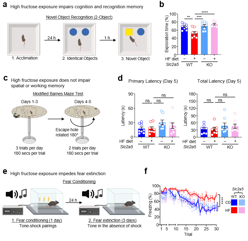
Impaired cognition/recognition memory: No preference for novel objects in the Novel Object Recognition (NOR) test (Fig. 4b).
Persistent fear responses: Profound fear extinction deficits (failure to suppress conditioned fear after threat removal)—a hallmark of anxiety disorders/PTSD (Fig. 4f).
■Rescue by GLUT5 deletion: GLUT5-KO mice on HF diet showed normal NOR performance (Fig. 4b) and intact fear extinction (Fig. 4f), proving GLUT5 mediates fructose-induced behavioral pathology.

Fig.4 Early-life high fructose exposure impairs juvenile behavioral adaptability
Significance and Implications
This study provides the first mechanistic explanation for fructose-associated neurodevelopmental risks:
GLUT5-dependent fructose uptake → Microglial metabolic rewiring (↑ F6P, oxidized state) → Impaired phagocytosis (synaptic pruning, efferocytosis) → Disrupted neurodevelopment → Cognitive/behavioral deficits (↑ Anxiety risk).
Key Takeaways
Critical Exposure Window: Pregnancy, lactation, and early childhood (synaptic pruning continues into adolescence in humans).
Primary Dietary Culprits: HFCS-sweetened beverages, juices, ultra-processed foods.
Public Health Action: Limit maternal/child intake of added sugars, especially liquid fructose.
Therapeutic Target: GLUT5 or downstream effectors (e.g., HK2) may offer intervention strategies.
Related Products List:
|
Catalog |
Product Name |
|
Recombinant Human SLC2A5 Protein, N-His-SUMO |
|
|
Anti-Rat SLC2A5 Antibody |
|
|
Anti-Human SLC2A5 Polyclonal Antibody |
